Product Information
| Product Name | Catalog No. | Specification | Storage | Shelf Life |
|---|---|---|---|---|
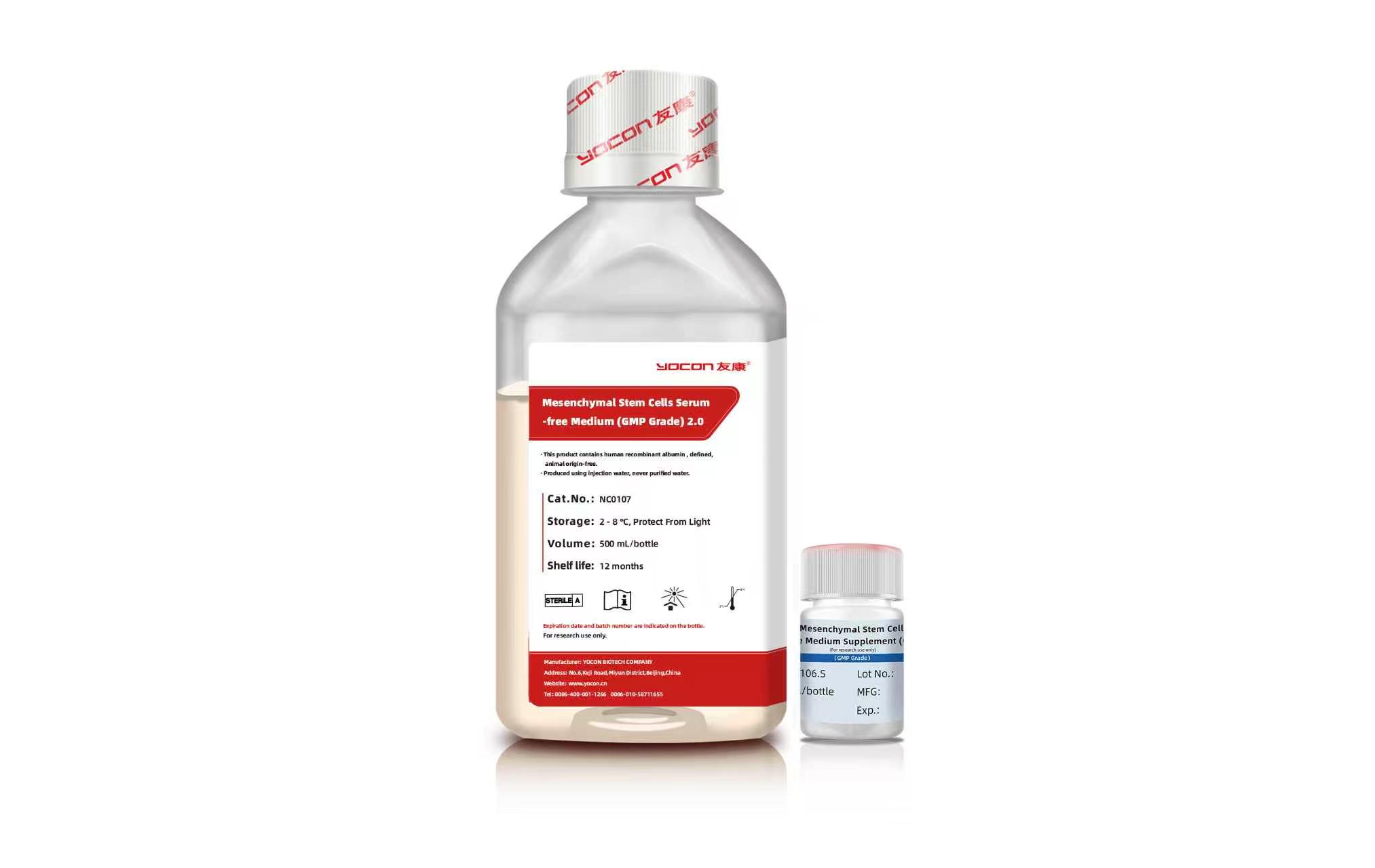
| Mesenchymal Stem Cells Serum-free Medium (GMP Grade) 2.0 | NC0107 | 500 mL/bottle | 2–8 ℃, protected from light | 12 months |
| Mesenchymal Stem Cells Serum-free Medium Supplement (GMP Grade) | NC0106.S | 5 mL/bottle | -20 ℃, protected from light | 12 months |
Product Advantages
Serum-free, defined cytokine culture system; no human or animal-derived materials; all proteins are recombinantly produced. Suitable for stem cell drug IND submission and clinical cell bank establishment, with FDA DMF filing (DMF No.: 31623).
Significantly improves cell adherence and adaptability, supports lower seeding density and higher expansion fold.
No medium exchange required, one-batch continuous culture; efficient passaging up to P20 without cellular senescence.
Compatible with cell factories, microcarriers, bioreactors and other 2D & 3D large-scale culture platforms.
Low background exosome content, facilitating downstream purification and enabling high-yield, high-quality exosome harvest.
Performance Data
1.Cell Performance
① Cell Passaging Performance
Umbilical cord primary MSC isolation and serial passaging
| Passage | Culture Vessel | Average Cell Yield per Vessel | Expansion Fold | Total Harvested Cells | Expansion Fold vs. P0 | Seeding Density /cm² | Culture Period |
|---|---|---|---|---|---|---|---|
| P0 | 150mm dish | 3.68E+06 | / | 2.2E7 | 1 | 35 tissue pieces /dish | 14–16 days |
| P1 | 1.50E+07 | 10.00 | 22 E7 | 10 | 8000 | 3 days | |
| P2 | 1.36E+07 | 11.29 | 249 E7 | 113 | |||
| P3 | 1.53E+07 | 12.75 | 3178 E7 | 1439 | |||
| P4 | 1.39E+07 | 11.55 | 36710 E7 | 16626 | |||
| P5 | 1.21E+07 | 10.08 | 370037 E7 | 167589 | |||
| P6 | 1.54E+07 | 10.28 | 3803985 E7 | 1722819 | 10000 | ||
| P7 | 1.75E+07 | 11.64 | 44278389 E7 | 20053618 | |||
| P8 | 1.50E+07 | 10.00 | 442783886 E7 | 200536180 | |||
| P9 | 1.73E+07 | 11.56 | 5119591718 E7 | 2318198242 | |||
| P10 | 1.45E+07 | 9.64 | 49343127758 E7 | 22347431050 |
② Cell Morphology

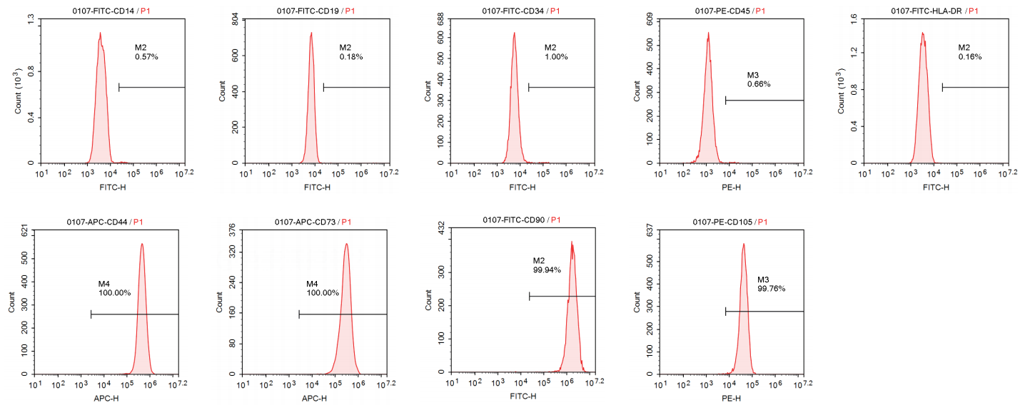
③ Surface Marker Characterization
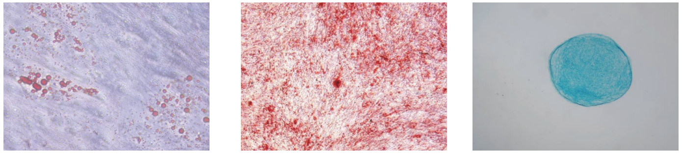
④ Trilineage Differentiation (P10 Cells)
⑤ Immunomodulatory Capacity
| Test Item | Test Method | Result |
|---|---|---|
| Lymphocyte Proliferation Inhibition Rate | Pharmacopoeia of the People's Republic of China (2020 Edition, Volume IV) General Chapter 3429, Immunochemistry, Flow Cytometry | 30.80% |
| Inhibition Rate of Lymphocyte TNF-α Secretion | Pharmacopoeia of the People's Republic of China (2020 Edition, Volume IV) General Chapter 3429, Immunochemistry, Flow Cytometry | 60.80% |
| Th1 Cell Proliferation Inhibition Rate | Pharmacopoeia of the People's Republic of China (2020 Edition, Volume IV) General Chapter 3429, Immunochemistry, Flow Cytometry | 63.60% |
| Th17 Cell Proliferation Inhibition Rate | Pharmacopoeia of the People's Republic of China (2020 Edition, Volume IV) General Chapter 3429, Immunochemistry, Flow Cytometry | 70.50% |
| Treg Cell Proliferation Promotion Rate | Pharmacopoeia of the People's Republic of China (2020 Edition, Volume IV) General Chapter 3429, Immunochemistry, Flow Cytometry | 31.60% |
⑥ Telomerase Activity
Test results of telomerase activity detected by RQ-TRAP assay
| Test Sample | FAM Channel Ct Value | Telomerase Activity (TPG/10000 cells) |
|---|---|---|
| Negative Control (Heat-inactivated Positive Control) | Undetermined | Not Detected |
| Heat-inactivated Sample Control | Undetermined | Not Detected |
| Positive Control | 22.7 | 3626.6 |
| NC0107-MSC (S250418-033) | 28.55 | 6.77 |
⑦ Chromosomal Karyotype Analysis
Statistical results of chromosomal karyotype analysis on metaphase cells
| Test Sample | Chromosome Analysis Item | Number of Abnormal Cells | Reference Value* |
|---|---|---|---|
| 250606 (S250609 - 030) | Chromatid and Chromosome Breaks | 0 | ≤8 |
| Structural Aberration | 0 | ≤2 | |
| Hyperploid | 2 | ≤2 | |
| Hypoploid | 6 | ≤18 | |
| Polyploid | 0 | ≤4 |
*Pharmacopoeia of the People's Republic of China, 2020 Edition Description of Results: G-banded karyotype: 46, XX. Karyotype analysis of 100 metaphase cells shows that chromatid and chromosome breaks, structural aberrations, hyperploidy and hypoploidy are all within the standard reference range for human diploid somatic cell chromosome analysis specified in Volume III of the Pharmacopoeia of the People's Republic of China (2020 Edition).
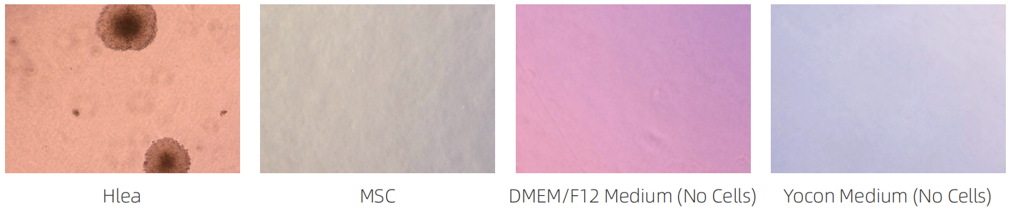
⑧ MSC In Vitro Tumorigenicity Test — Soft Agar Colony Formation Assay
After 21 days of culture, HeLa cells formed abundant cell colonies, while no cell colonies were observed in MSC groups. MSCs exhibit no tumorigenicity in in vitro culture.
2. Exosome Culture Performance
① Exosome Culture Cases
Case 1: This product is used for exosome production in Cell Factory P2 cells were recovered, continuously passed to P3 and seeded into cell factories. A total of 2.52E+12 exosomes were harvested within 15 days.
Calculation of Total Harvested Exosomes
| Sampling Time Point | Exosome Concentration (particles/mL) | Harvested Exosome Quantity (particles) | Medium Volume (mL) |
|---|---|---|---|
| Day 3 | 3.53E+09 | 4.24E+11 | 120 |
| Day 6 | 5.36E+09 | 6.43E+11 | 120 |
| Day 9 | 4.76E+09 | 5.71E+11 | 120 |
| Day 12 | 5.46E+09 | 6.55E+11 | 120 |
| Day 15 | 1.91E+09 | 2.29E+11 | 120 |
| Total | — | 2.52E+12 | 600 |
Case 2: This product is applied for exosome harvest from microcarrier-based 3D cell culture Culture cycle: 15 days, 500 mL medium; total harvested exosomes: 3.50E+12 particles.
Calculation of Total Harvested Exosomes
| Sampling Time Point | Exosome Concentration (particles/mL) | Harvested Exosome Quantity (particles) | Medium Volume (mL) |
|---|---|---|---|
| Day 3 | 6.19E+09 | 6.19E+11 | 100 |
| Day 6 | 9.26E+09 | 9.26E+11 | 100 |
| Day 9 | 7.92E+09 | 7.92E+11 | 100 |
| Day 12 | 6.06E+09 | 6.06E+11 | 100 |
| Day 15 | 5.54E+09 | 5.54E+11 | 100 |
| Total | — | 3.50E+12 | 500 |
Case 3: This product is used for large-scale exosome production on Yocon Exosome Culture Workstation One culture cycle lasts 15 days with 5 L medium, harvesting 1 L supernatant every 3 days. The total harvested exosome quantity reaches 2.34E+13 particles.
Calculation of Total Harvested Exosomes
| Sampling Time Point | Exosome Concentration (particles/mL) | Harvested Exosome Quantity (particles) | Medium Volume (mL) |
|---|---|---|---|
| Day 3 | 4.74E+09 | 4.74E+12 | 1000 |
| Day 6 | 5.93E+09 | 5.93E+12 | 1000 |
| Day 9 | 4.24E+09 | 4.24E+12 | 1000 |
| Day 12 | 4.22E+09 | 4.22E+12 | 1000 |
| Day 15 | 4.30E+09 | 4.30E+12 | 1000 |
| Total | — | 2.34E+13 | 5000 |
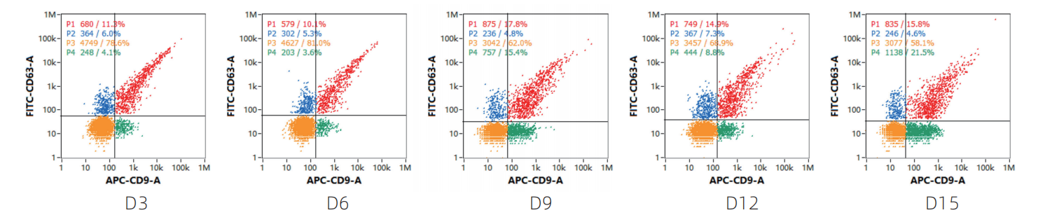
② Nanoflow Detection of Exosome Surface Protein Markers (CD9 and CD63)
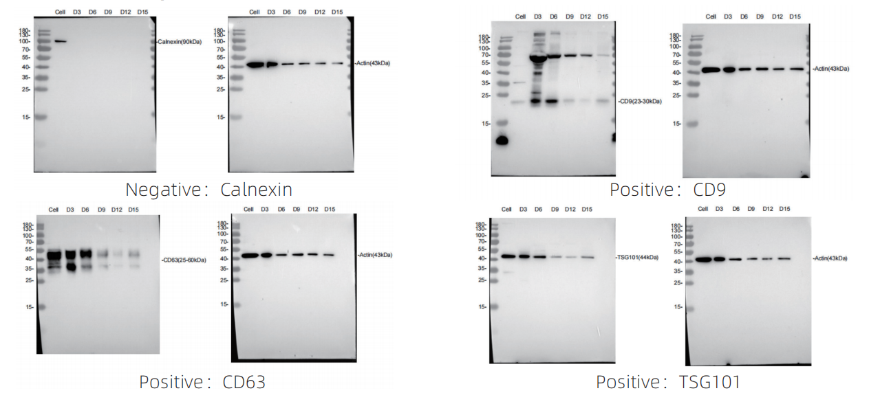
③ Western Blot (WB) Detection of Protein Markers (Negative Marker: Calnexin; Positive Markers: CD9, TSG101 and CD63)